Valve Function Weakens
One-way valves may stop keeping blood moving upward efficiently.

 EN
Texas Leg Skin & Vein Evaluation
Color changes on the legs can come from the skin itself, an injury, diabetes, infection, arteries, veins, or other medical conditions. Texas Vein Experts looks for a venous pattern only when the location, texture, swelling, visible veins, and other findings make vein disease a reasonable possibility.
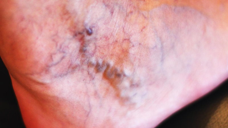
Brown, red & purple patterns
Ultrasound when indicated

Start With What You Can See
The most useful first questions are where the color change appears, whether it is new or persistent, what the skin feels like, and whether swelling or other leg symptoms occur with it.
Color
Brown, rust, red, purple, blue, pale, or nearly black changes can point toward different processes.
Location
Venous skin changes often involve the lower leg or ankle, while other causes may appear in different patterns.
Texture and symptoms
Itching, scaling, thickening, warmth, swelling, pain, visible veins, or an open sore can change what type of evaluation is needed.
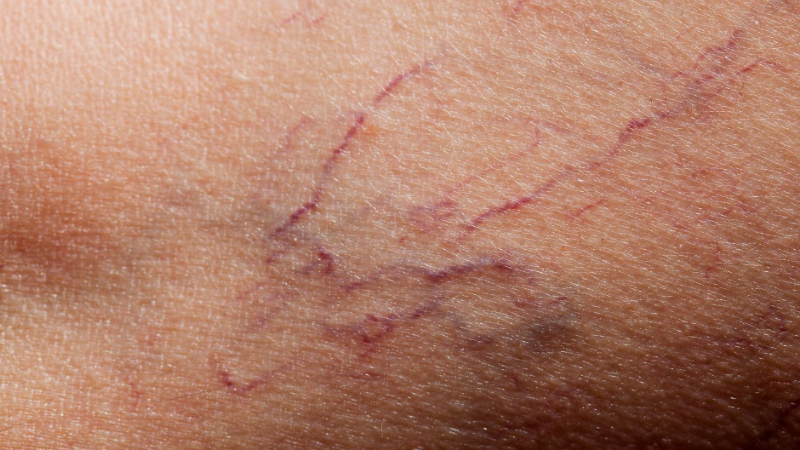
One Venous Pathway
When discoloration is caused by chronic venous disease, the color change can develop gradually through a chain of pressure, leakage, inflammation, and pigment deposition in the lower-leg tissues.
One-way valves may stop keeping blood moving upward efficiently.
Backward flow can increase pressure in veins and small vessels of the lower leg.
Persistent pressure can allow fluid and red-blood-cell components to enter nearby tissue.
Breakdown of red blood cells can leave hemosiderin, an iron-containing pigment, in the tissue.
Brown or rust-colored staining can appear with dryness, inflammation, or tissue thickening over time.
Possible Venous Clues
No single color confirms venous disease. These associated findings make a venous explanation more plausible, especially when they cluster around the ankle or lower leg.
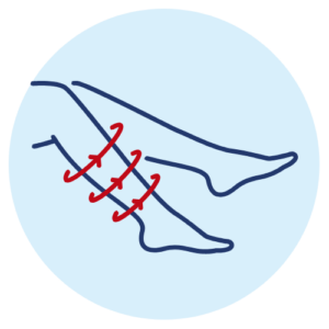
Persistent reddish-brown or darker staining can develop near the ankle or lower leg.
Swelling that becomes more noticeable later in the day can accompany chronic venous pressure.
Venous stasis dermatitis can make the skin irritated, flaky, itchy, or inflamed.
Long-standing inflammation may make the lower-leg skin and underlying tissue feel harder or tighter.
Visible bulging veins or a heavy, aching feeling can strengthen the venous pattern.

Open or slow-healing areas require medical assessment and may indicate more advanced tissue changes.
Color is only one clue. Location, swelling, skin texture, visible veins, temperature, pain, and how quickly the change appeared help determine what type of evaluation makes sense.
Explore Related Symptoms
Possible Causes
The same brown, red, purple, or darkened appearance can result from very different conditions. The underlying cause determines whether vein care, dermatology, urgent care, or another medical evaluation is appropriate.
Venous hypertension and stasis dermatitis can cause brown or reddish changes, swelling, itching, and tissue thickening.
Eczema, psoriasis, contact dermatitis, and other inflammatory skin disorders can change both color and texture.
Bruises, bites, shaving irritation, cuts, or prior inflammation can leave temporary or persistent darker patches.
Diabetic dermopathy and other metabolic skin findings can cause brown or scaly patches on the lower legs.
Reduced arterial flow can cause pale, blue, cool, or slow-healing skin changes and requires a different vascular evaluation.
Cellulitis, DVT, medication effects, vasculitis, and other conditions can cause new redness, purple change, swelling, or tenderness.
Compare the Clues
These patterns are not a self-diagnosis tool. They show why the location, texture, temperature, timing, and associated symptoms matter as much as the color itself.
Possible Venous Pattern
More suggestive when the discoloration sits near the ankle and occurs with swelling, aching, heaviness, itching, skin thickening, or varicose veins.
Skin / Pigment Pattern
Rashes, sun-related pigment, irritation, bruising, or healing inflammation can darken the skin without a venous reflux pattern.
Different / Urgent Vascular Pattern
Reduced arterial flow, a clot, or another acute problem can produce a very different pattern from chronic venous staining.
Know When to Act
Persistent or progressive color change deserves attention when the cause is unclear, especially if the skin is changing in texture or the discoloration occurs with swelling, pain, visible veins, or a wound.
Some sudden color changes need prompt medical assessment rather than a routine vein appointment.
The Discoloration Persists or Becomes Darker
A mark that does not fade like a normal bruise, spreads, or changes over time should be evaluated rather than judged by color alone.
It Is Centered Around the Ankle or Lower Leg With Venous Symptoms
Brown or rust-colored skin plus swelling, heaviness, aching, itching, or varicose veins can fit a chronic venous pattern.
The Skin Is Itchy, Scaly, Thickened or Firm
Changes in both color and texture may reflect dermatitis, chronic inflammation, or another skin process that deserves assessment.
A Sore Opens or Is Slow to Heal
Skin breakdown or a wound around the lower leg or ankle needs medical attention and evaluation of the underlying cause.
The Change Is Sudden or Comes With Concerning Symptoms
Sudden one-sided swelling, pain, warmth, or redness needs prompt medical assessment. A suddenly cold, pale or blue, numb, or weak foot can be an arterial emergency.
Diagnosis Before Vein Treatment
Texas Vein Experts does not assume that discoloration is caused by veins. The goal is to decide whether the history, examination, and—when appropriate—duplex ultrasound support a venous explanation.
Discuss when the discoloration started, whether it is changing, where it appears, prior injuries or clots, and symptoms such as itching, swelling, pain, or heaviness.
Look at color, texture, swelling, visible veins, tenderness, asymmetry, and other findings that may point toward or away from a venous cause.
If venous disease is suspected, ultrasound can assess blood flow, valve function, reflux, and which superficial veins are involved.
If reflux is confirmed, discuss appropriate vein care. If the pattern is dermatologic, arterial, infectious, or otherwise non-venous, another evaluation may be more appropriate.
Treatment for Confirmed Venous Reflux
When a venous condition is actually contributing to the lower-leg changes, Texas Vein Experts may recommend one or more minimally invasive treatments based on the refluxing vein and anatomy. Existing pigmentation may improve slowly, remain visible, or require separate skin care.
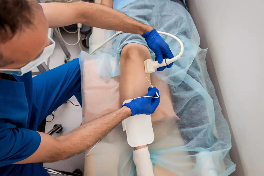
Thermal Catheter Treatment
Controlled radiofrequency energy closes an eligible refluxing superficial source vein under ultrasound guidance.
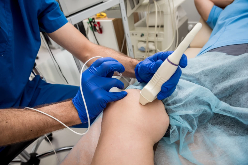
Prescription Microfoam
Polidocanol microfoam can treat selected refluxing superficial veins and associated varicosities when the anatomy is appropriate.
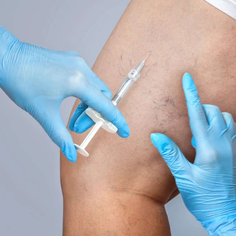
Injection Treatment
A physician-prepared sclerosant may treat selected smaller varicose, reticular, or residual surface veins as part of an individualized plan.
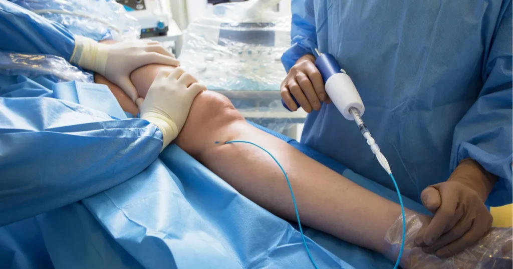
Nonthermal Medical Adhesive
A catheter delivers medical adhesive to close an eligible refluxing source vein without thermal energy along the treated segment.
Important: treating venous reflux does not guarantee that existing brown or dark pigmentation will disappear. Skin color can improve slowly, remain visible, or need separate dermatologic management depending on the cause and duration of the change.
If a Vein Problem Is Confirmed
The procedure is selected for the diagnosed venous anatomy. Skin appearance can change on a different timeline from swelling, aching, or other vein-related symptoms.
01
Review the Diagnosis and Target Vein
Confirm the clinical findings, duplex results, procedure, medications, and preparation instructions.
Prepare the Treatment Area
The leg is cleaned and prepared. Comfort measures depend on the selected procedure.
Treat the Diagnosed Venous Problem
The physician treats the selected refluxing or surface veins rather than treating discoloration as a standalone diagnosis.
02
Follow Procedure-Specific Aftercare
Walking, compression, bathing, travel, and activity guidance can differ by treatment and patient.
Protect and Monitor the Skin
Continue any skin-care or wound-care instructions and report new redness, warmth, drainage, or skin breakdown.
Reassess Symptoms and Skin Changes
Follow-up focuses on healing, venous symptoms, remaining veins, and whether the skin findings need additional care.
Medical Necessity & Benefits
Skin pigmentation or eczema related to chronic venous insufficiency can be clinically important, but coverage depends on the diagnosed venous condition, examination, ultrasound findings, plan criteria, and medical-necessity requirements. Discoloration alone does not guarantee coverage.
Clinical Evaluation
Document the skin changes, associated symptoms, prior care, and examination findings.
Venous Imaging When Appropriate
Use duplex ultrasound to document reflux and vein anatomy when a venous cause is suspected.
Benefits and Plan Requirements
Review referrals, authorization, documentation, conservative-care requirements, and medical-necessity criteria.
Clear Financial Next Steps
Explain the approved pathway and estimated patient responsibility before a procedure is scheduled.
Why Texas Vein Experts?
We do not diagnose from color alone
The location, texture, swelling, symptoms, history, and physical findings all matter before the discoloration is connected to vein disease.
Duplex ultrasound is used when the venous pattern fits
Imaging can show whether reflux is actually present and which superficial veins are involved.
Four minimally invasive options support individualized care
RFA, Varithena, compounded sclerotherapy, and VenaSeal can serve different roles when a treatable venous condition is confirmed.
Physician-led care across three Texas regions
The TVE physicians serve clinics throughout DFW, Houston, and the San Antonio area, with benefits review before treatment.
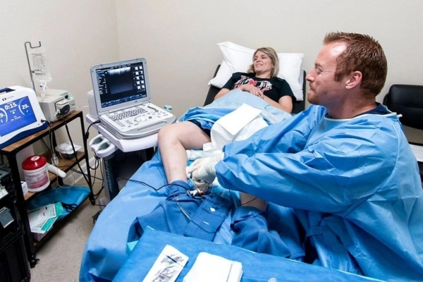
Your Texas Vein Care Team
When discoloration may be connected to chronic venous disease, TVE physicians combine the clinical picture with venous imaging when appropriate before recommending treatment.
Our Locations
Find a Texas Vein Experts clinic near you in Dallas–Fort Worth, Houston or San Antonio. Open a region to see every location.

Dallas–Fort Worth
9 clinics
Houston
6 clinics
San Antonio
4 clinicsCommon Questions
Clear answers to common patient questions about possible causes, when to get checked, vein evaluation, and treatment options.
Possible causes include sun exposure, bruising, post-inflammatory hyperpigmentation, eczema and other skin conditions, diabetes-related changes, infection, medications, arterial problems, chronic venous disease, and other medical conditions. The appearance alone does not establish the cause.
Persistent brown or rust-colored ankle changes can occur with chronic venous pressure, especially when swelling, heaviness, itching, skin thickening, or varicose veins are also present. Other causes are possible, so the pattern should be evaluated rather than assumed.
Hemosiderin is an iron-containing pigment that can remain in tissue after red blood cells break down. In chronic venous disease, prolonged pressure and leakage from small vessels can contribute to brown or rust-colored staining in the lower legs.
Yes. Chronic venous insufficiency can cause lower-leg swelling, inflammation, reddish-brown pigmentation, itching, scaling, and tissue changes. Arterial circulation problems can also change skin color, but they follow a different pattern and require different testing.
Purple or blue color can have several causes, including bruising, cold-related vessel changes, venous congestion, livedo patterns, blood clots, or reduced arterial flow. A sudden cold, pale or blue, numb, or weak foot needs urgent medical assessment.
Yes. Diabetes can be associated with skin findings such as diabetic dermopathy and may also affect wound healing and circulation. A vein clinic should not assume that discoloration in a person with diabetes is venous without an appropriate evaluation.
A venous pattern often involves persistent brown or reddish-brown changes near the ankle or lower leg and may occur with swelling, itching, scaling, skin thickening, aching, heaviness, or visible varicose veins.
Seek medical evaluation for persistent or progressive changes, skin thickening, significant swelling, pain, an open sore, or changing skin lesions. Sudden one-sided swelling with pain, warmth, or redness needs prompt assessment. A suddenly cold, pale or blue, numb, or weak foot can be an emergency.
Not necessarily. Brown pigmentation has many possible causes. Chronic venous disease becomes more plausible when the discoloration is concentrated near the lower leg or ankle and occurs with swelling, heaviness, itching, varicose veins, or other venous findings.
Not everyone with skin discoloration needs a venous ultrasound. When the history and physical findings suggest possible venous disease, duplex ultrasound can evaluate blood flow, valve function, reflux, and the veins involved.
Topical products may help dryness, irritation, or certain pigment conditions, but they do not correct underlying venous reflux. Existing pigmentation caused by chronic venous disease can be difficult to reverse and may need separate dermatologic care.
Not necessarily. Treating a confirmed venous problem addresses abnormal vein function, not pigment directly. Some skin changes may improve gradually, while established staining or tissue changes can remain visible.
TVE offers Radiofrequency Ablation, Varithena, Compounded Sclerotherapy, and VenaSeal for selected venous conditions. The appropriate option depends on the diagnosis, vein anatomy, ultrasound findings, and individual medical considerations.
Yes. More advanced chronic venous disease can include pigmentation, skin thickening, inflammation, and venous ulcers. Arterial disease, diabetes, infection, and other conditions can also contribute to wounds, so a non-healing sore needs medical evaluation.
Coverage depends on the diagnosed venous condition, clinical findings, ultrasound results, medical necessity, prior authorization, and the requirements of the individual insurance plan. Skin discoloration by itself does not guarantee coverage.
 English English
 Spanish