Small Surface Vessels Dilate
Tiny vessels close to the skin can widen and become visible as red, blue, or purple lines.

 EN
Texas Spider Vein Care
Spider veins are small red, blue, or purple surface vessels that often form web-like or branching patterns. They are usually an appearance concern rather than a dangerous condition, but the surrounding vein pattern and any leg symptoms can change what evaluation or treatment makes sense.
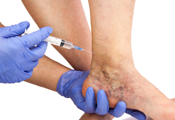
Spider-vein pattern review
Sclerotherapy when appropriate

Start With What You Can See
Spider veins, also called telangiectasias, are very small visible blood vessels near the skin surface. On the legs they often look like fine red, blue, or purple lines arranged in webs, branches, or small clusters.
They sit close to the skin
Spider veins are much smaller and flatter than bulging varicose veins and usually do not raise the skin.
They may connect with reticular “feeder” veins
Slightly larger blue or green reticular veins can sit beneath a spider-vein network and may influence how it is treated.
They are usually an appearance concern
Most isolated spider veins do not cause significant symptoms or require treatment for medical reasons.

Why They Appear
Spider veins can look similar even when the reason behind them is different. Unlike large varicose veins, isolated spider veins are not automatically proof of chronic venous insufficiency.
Tiny vessels close to the skin can widen and become visible as red, blue, or purple lines.
Family history, pregnancy, and hormonal changes can make spider veins more likely to develop.
Long periods of standing or sitting, aging, weight, or prior local injury can influence visible leg vessels.
Some spider-vein networks sit over reticular veins or coexist with deeper venous reflux, especially when other leg symptoms are present.
What You May Notice
For many people, spider veins are visible without pain, swelling, or other leg symptoms. The appearance itself is often the main concern.
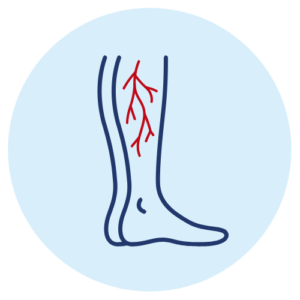
Fine vessels may appear as red, blue, or purple markings just beneath the skin.
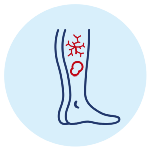
The pattern can resemble a spider web, tiny tree branches, or a sunburst.
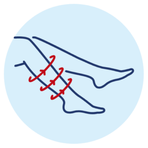
Spider veins usually remain flat rather than forming rope-like or bulging veins.
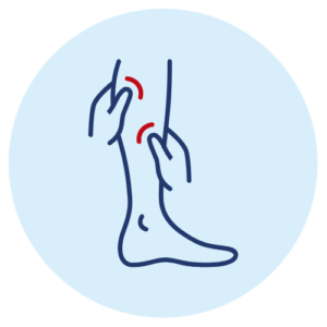
Leg spider veins may appear in a small patch or across a broader area.

Isolated spider veins frequently do not cause aching, swelling, or heaviness.
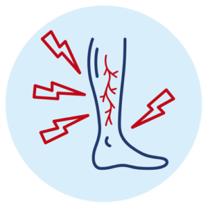
Some people notice localized burning, itching, or tenderness around a spider-vein network.
Symptoms change the question. If spider veins appear with recurring swelling, heaviness, aching, skin changes, or larger varicose veins, the evaluation should look beyond the surface pattern.
Explore Related Symptoms
Know the Difference
Size, depth, and whether a vessel bulges above the skin help distinguish the visible vein types and guide the treatment approach.
Fine / Web-Like
Very small red, blue, or purple surface vessels that usually remain flat.
Blue / Green Feeder Veins
Slightly larger blue or green veins beneath the skin that can feed nearby spider-vein networks.
Raised / Bulging
Larger superficial veins that may twist, bulge, and occur with aching, heaviness, or swelling.
Cosmetic Concern or Something More?
You do not have to treat spider veins simply because they are visible. An evaluation can still be useful when you want them reduced or when the surrounding symptom pattern raises a different question.
The Appearance Bothers You
Cosmetic concern alone is a valid reason to ask about treatment, even when there are no other symptoms.
Burning, Itching, or Tenderness Keeps Returning
Persistent local symptoms deserve assessment rather than assuming the visible spider veins are the only cause.
You Also Have Heaviness, Swelling, or Bulging Veins
Those findings point beyond isolated spider veins and may justify evaluation for a larger venous problem.
There Are Dense Veins Around the Ankle With Skin Changes
An ankle flare, discoloration, rash, or other lower-leg skin change deserves a broader venous assessment.
A Vein Suddenly Becomes Red, Warm, Swollen, or Very Tender
Prompt medical evaluation is appropriate when a visible vessel changes suddenly or significant new leg symptoms appear.
Diagnosis Before Treatment
For isolated spider veins without significant symptoms, routine venous ultrasound is usually unnecessary. Texas Vein Experts can use imaging selectively when the history or examination suggests a deeper refluxing vein, recurrent disease, or another venous concern.
Start by identifying whether the main concern is appearance, local discomfort, or a broader leg-symptom pattern.
Look at spider veins, reticular feeder veins, bulging varicose veins, skin changes, and where the vessels are located.
Recurring swelling, heaviness, significant burning or pain, bleeding, or larger veins can change the diagnostic plan.
If the clinical picture suggests underlying venous incompetence, ultrasound can map reflux and help determine whether a larger source vein should be addressed first.
Personalized Spider Vein Care
Compounded sclerotherapy can directly treat selected leg spider veins. The other TVE procedures below are not routine spider-vein treatments; they become relevant only when the evaluation identifies a larger refluxing or varicose vein that should be treated as part of the overall plan.
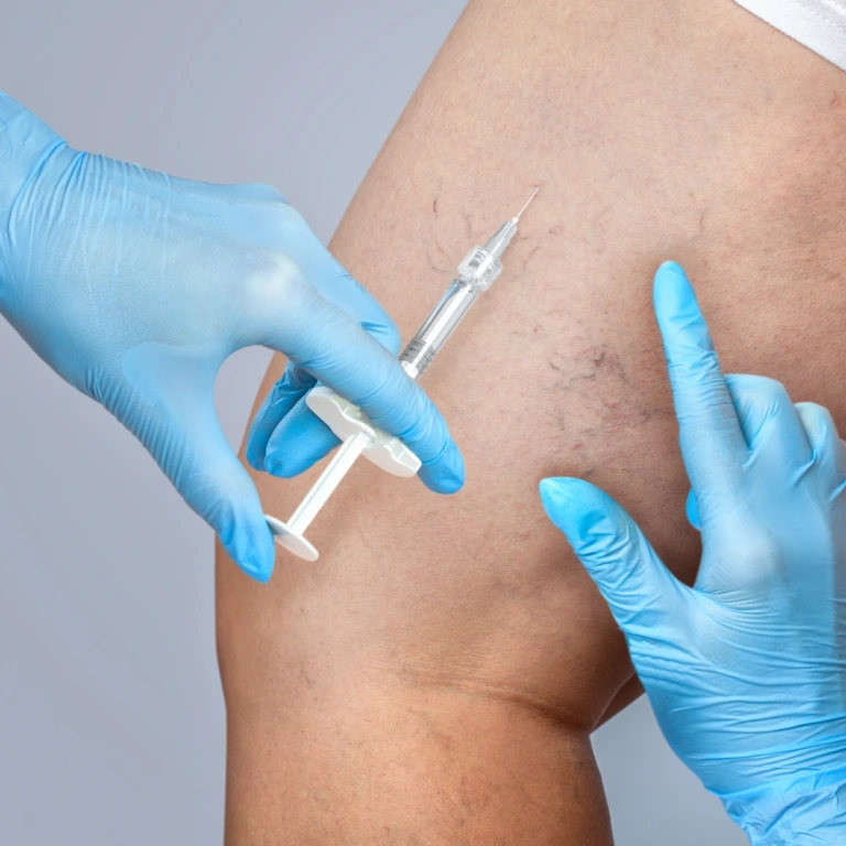
Direct Spider Vein Treatment
A clinician injects a selected sclerosant into targeted spider or reticular veins so the vessels close and gradually become less visible.
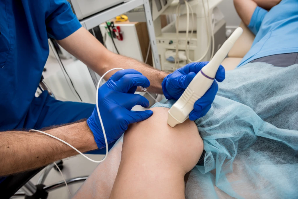
Prescription Microfoam
Varithena is used for eligible refluxing superficial veins and associated varicosities. It is not a routine treatment for isolated spider veins.
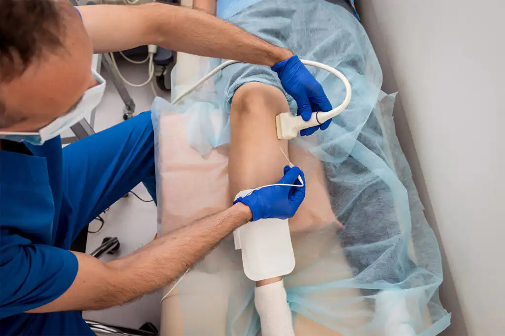
For Diagnosed Source Reflux
RFA closes an eligible refluxing superficial source vein with controlled radiofrequency energy. It does not directly erase the small spider-vein network on the skin.
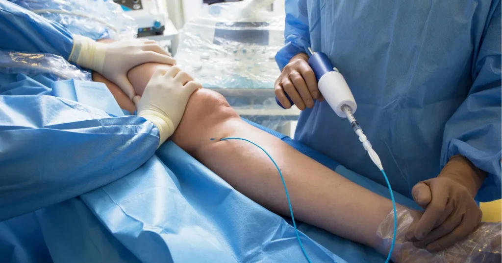
For Diagnosed Source Reflux
VenaSeal uses medical adhesive to close an eligible refluxing superficial vein. It is considered only when a larger source vein, not the spider veins themselves, requires treatment.
The treatment should match the vein type. Isolated spider veins and a refluxing saphenous vein are different problems, so the same procedure should not be used for every visible vein.
Before, During & After Treatment
The details depend on whether the plan is limited to surface sclerotherapy or includes treatment of a larger underlying vein first. Expectations should be set around gradual fading, not an instant cosmetic result.
01
Confirm the Veins and the Goal
The clinician reviews the spider-vein pattern, any feeder veins, and whether the plan is cosmetic surface treatment or part of broader venous care.
Prepare the Treatment Area
The skin is cleaned and the treatment area is marked or examined closely. Comfort measures depend on the selected treatment.
Treat the Planned Veins
For direct spider-vein treatment, small injections are placed into selected surface vessels. If a deeper source vein is being treated first, the procedure follows that separate plan.
02
Expect Temporary Skin Changes
Redness, bruising, mild tenderness, firmness, or temporary pigmentation can occur as treated vessels close and heal.
Follow Walking, Compression, and Skin-Care Instructions
Aftercare varies according to the veins treated and the procedure used. Follow the individualized instructions provided by the clinical team.
Allow Time for Fading and Reassessment
Spider veins usually fade gradually. Multiple sessions may be needed, and new spider veins can develop later even after successful treatment.
Why Texas Vein Experts?
We do not assume every spider vein means chronic venous insufficiency
Isolated spider veins are often cosmetic. The evaluation looks for symptoms and larger vein findings before deciding whether deeper testing is useful.
Compounded sclerotherapy directly targets selected surface veins
For many leg spider-vein patterns, targeted injection treatment is the most relevant TVE procedure.
Ultrasound is used selectively when the clinical picture calls for it
Significant symptoms, larger varicose veins, skin changes, bleeding, or recurrent/complex patterns can justify a deeper venous assessment.
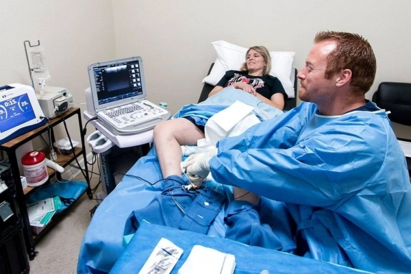
Four treatment options are available when different vein levels are involved
Sclerotherapy treats surface veins, while Varithena, RFA, or VenaSeal may have a role only when a larger refluxing vein is diagnosed and needs treatment first.
Your Texas Vein Care Team
These TVE physicians evaluate visible vein patterns and can determine whether the concern is limited to surface spider veins or connected to a broader venous condition that changes the treatment plan.
Our Locations
Find a Texas Vein Experts clinic near you in Dallas–Fort Worth, Houston or San Antonio. Open a region to see every location.

Dallas–Fort Worth
9 clinics
Houston
6 clinics
San Antonio
4 clinicsCommon Questions
Clear answers to common patient questions about possible causes, when to get checked, vein evaluation, and treatment options.
Spider veins, also called telangiectasias, are very small visible blood vessels near the skin surface. They commonly appear as flat red, blue, or purple lines in web-like, branch-like, or sunburst patterns.
Spider veins can develop for several reasons, including genetics, pregnancy and hormonal changes, aging, prolonged standing or sitting, local injury, and other vessel changes. In some people they coexist with reticular veins or deeper venous reflux, but they are not automatically caused by chronic venous insufficiency.
Not necessarily. Isolated spider veins are often a cosmetic finding. If they occur with swelling, heaviness, aching, skin changes, or larger varicose veins, an evaluation may be useful to determine whether a deeper venous problem is present.
No. Treatment is optional when spider veins are isolated and do not cause symptoms. Some people choose treatment because the appearance bothers them, while others seek evaluation because of local discomfort or additional leg symptoms.
Professional treatment is usually needed to make established spider veins fade. For selected leg spider veins, sclerotherapy is a common treatment because it closes the targeted vessels so they gradually become less visible.
Lifestyle measures may support general vein health or reduce the chance of new veins becoming noticeable, but they do not reliably eliminate an established spider-vein network. Be cautious with products that promise to erase spider veins without a procedure.
Spider veins are much smaller, flatter, and closer to the skin surface. Varicose veins are larger and may twist or bulge above the skin. Reticular veins are medium-size blue or green vessels that can sit between the two and sometimes feed spider-vein networks.
Not everyone does. Current venous guidelines advise against routine duplex ultrasound for isolated asymptomatic spider veins. Ultrasound may be appropriate when significant symptoms, bleeding, larger varicose veins, skin changes, recurrent disease, or another clinical clue suggests underlying venous incompetence.
A clinician injects a sclerosant into selected spider or reticular veins. The medication irritates the vessel lining, causing the treated vessel to close and gradually fade as the body redirects blood through other functioning vessels.
The number of sessions varies with the size, number, distribution, and response of the treated veins. Spider-vein treatment is often staged, so one area may need more than one session before the cosmetic result is complete.
A successfully treated vessel may fade, but treatment does not prevent every future spider vein from developing. New vessels can appear over time because of genetics, hormones, aging, local pressure, or an untreated feeder or refluxing vein when one is present.
Yes. Pregnancy-related hormonal changes, increased blood volume, and pressure on pelvic and leg veins can make spider veins more noticeable. Some pregnancy-related vessels may improve after delivery, while others persist.
No. RFA and VenaSeal are designed for eligible larger refluxing superficial veins. They may have a role only when an evaluation identifies an underlying source vein that should be treated before or separately from the visible spider-vein network.
Varithena treats eligible refluxing superficial veins and associated varicosities rather than isolated spider veins. It may become relevant when the visible spider-vein concern is part of a larger venous-reflux pattern identified during evaluation.
Treatment performed only to improve the appearance of isolated spider veins is commonly considered cosmetic and may not be covered. If a separate medically necessary venous condition is diagnosed, coverage for that treatment depends on the individual plan, documentation, and authorization requirements.
 English English
 Spanish