Valve Closure Weakens
A one-way valve may no longer seal tightly enough to keep blood moving in one direction.

 EN
Texas Varicose Vein Care
Raised or twisted leg veins can be an appearance concern, a source of aching or swelling, or a clue that blood is moving backward through superficial veins. Texas Vein Experts looks at the pattern, your symptoms, and the venous circulation before deciding whether treatment is appropriate.
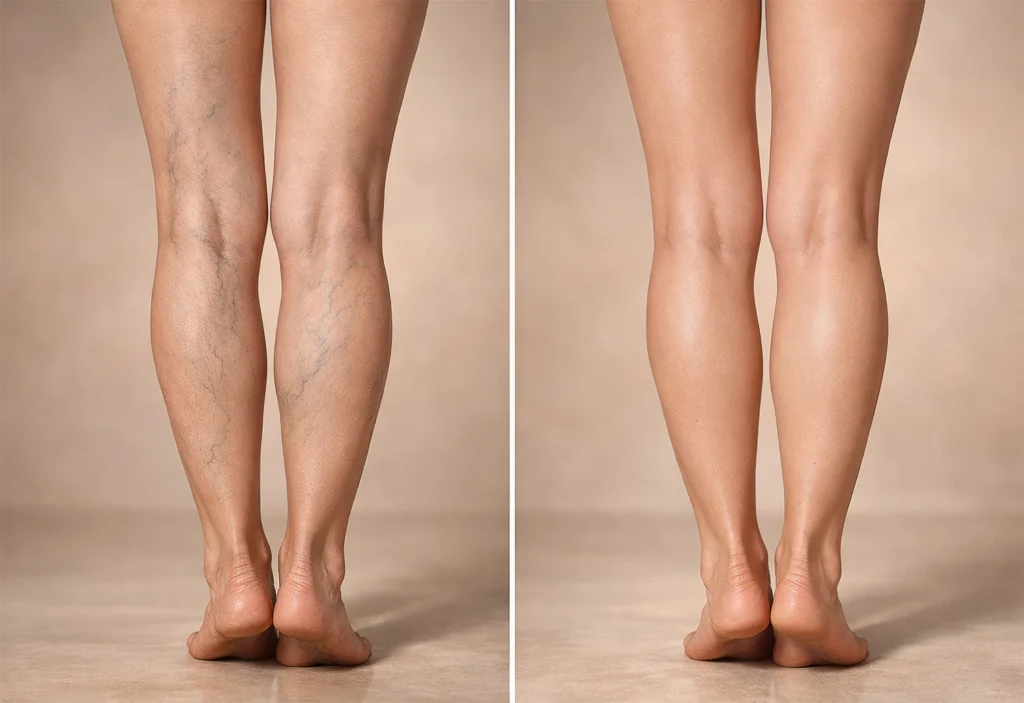
Ultrasound-guided planning
Four treatment pathways
Start With the Vein Itself
Leg veins carry blood back toward the heart. Small one-way valves help keep that flow moving upward. When a superficial vein stretches or a valve stops closing effectively, some blood can move backward and collect in the vein.
Pressure rises inside the superficial vein
The vein may widen as blood pools instead of moving efficiently upward.
The vein can lengthen and twist
As the wall stretches, the vein may become rope-like, raised, or bulging beneath the skin.
Appearance does not tell the whole story
Some varicose veins are mainly cosmetic. Others occur with aching, heaviness, swelling, itching, or more advanced skin changes.
How the Vein Changes
This is one common pathway. The exact source and extent of reflux can vary from one leg—and one patient—to another.
A one-way valve may no longer seal tightly enough to keep blood moving in one direction.
Backward flow, called venous reflux, lets more blood collect in the superficial vein.
Higher pressure can widen the vein and make the vessel wall more tortuous over time.
The enlarged vein may appear raised, twisted, or rope-like and may or may not cause symptoms.
What You May Notice
Some people are bothered mainly by appearance. Others develop a repeating pattern of leg symptoms, especially after long periods on their feet or sitting still.
A pressured, tired, or aching feeling that may become more noticeable as the day goes on.
Recurring swelling can occur when venous pressure is elevated, although swelling has many other possible causes.
Some symptomatic varicose veins are associated with throbbing, tightness, or cramping sensations.
Skin near a prominent vein may feel itchy, warm, or irritated, particularly around the lower leg.
An enlarged vein can feel sore or sensitive, especially along a bulging surface branch.
Persistent discoloration, irritation, or hardening can signal more advanced chronic venous disease and deserves evaluation.
Symptoms are clues, not a diagnosis. Aching, swelling, burning, cramping, and skin changes can come from conditions unrelated to the veins. The pattern and clinical findings matter.
Explore Related Symptoms
Visible Vein Types
Size, depth, shape, and the source of the vein all matter. A treatment that fits one visible-vein pattern may not fit another.
Raised / Bulging
Larger superficial veins that may be raised, twisted, or rope-like.
Blue / Green Feeder Veins
Medium-size blue or green veins that usually sit flatter beneath the skin.
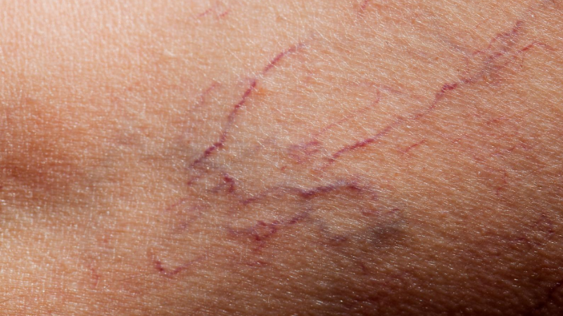
Small / Web-Like
Fine red, blue, or purple surface vessels that form small webs or branches.
Cosmetic or Medical?
There is no rule that every visible varicose vein must be treated. Evaluation becomes more useful when symptoms repeat, daily activity is affected, or the surrounding skin begins to change.
Symptoms keep returning
Persistent aching, heaviness, throbbing, itching, or tenderness can justify a closer look at the venous circulation.
Swelling follows a consistent pattern
Recurring lower-leg or ankle swelling—especially with visible veins—may make a venous evaluation more relevant.
The skin around the ankle is changing
Ongoing pigmentation, irritation, or hardening can occur with more advanced chronic venous disease.
A surface vein bleeds or a wound is slow to heal
Bleeding from a prominent vein or a persistent lower-leg wound deserves medical review rather than cosmetic self-treatment.
One leg changes suddenly
Sudden swelling, pain, warmth, or redness/discoloration needs prompt medical assessment because causes other than ordinary varicose veins must be considered.
Diagnosis Before Treatment
Texas Vein Experts uses the history, examination, and duplex ultrasound when appropriate to understand where abnormal flow begins and which veins are actually involved.
Discuss when symptoms occur, which leg is affected, how long changes have been present, and what matters most to you.
Review the distribution of bulging veins along with swelling, skin changes, tenderness, and other clinical findings.
Evaluate flow direction, valve function, reflux, vein anatomy, and other findings that can change the treatment plan.
Decide whether treatment is needed at all and, if so, which source vein and visible branches should be addressed first.
Personalized Vein Treatment
When ultrasound findings and the clinical picture confirm a treatable vein problem, Texas Vein Experts may recommend one or more minimally invasive options based on the diagnosed vein pattern.
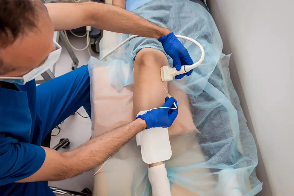
Thermal Catheter Treatment
A catheter delivers controlled radiofrequency energy to close an eligible refluxing superficial vein under ultrasound guidance.
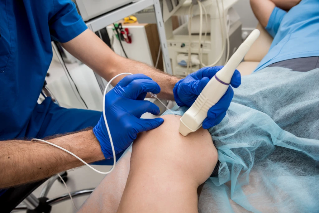
Prescription Microfoam
Polidocanol microfoam can be used for selected refluxing superficial veins and varicosities when the vein pattern is appropriate.
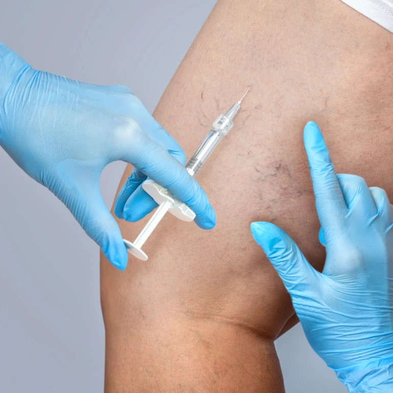
Targeted Injection Treatment
A liquid or foam sclerosant may be used for selected smaller varicose veins, reticular veins, spider veins, or residual surface branches.
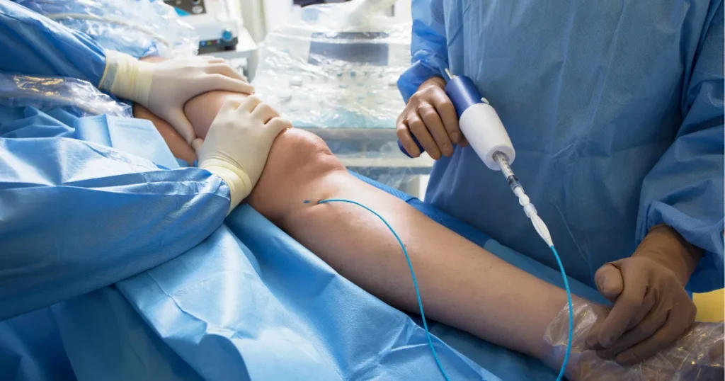
Nonthermal Medical Adhesive
A catheter delivers medical adhesive to close an eligible refluxing vein without using thermal energy along the treated segment.
One patient can need more than one technique. Treating the refluxing source vein and treating remaining visible branches are sometimes separate steps, so the plan may be staged rather than completed with one universal procedure.
Planning Through Follow-Up
Once the diagnosis is clear, the next step is understanding timing, preparation, and what recovery typically looks like after office-based treatment.
01
Reconfirm the ultrasound map
The team reviews the target vein, treatment sequence, and relevant medical information before starting.
Prepare for the selected technique
Comfort measures and preparation differ among thermal, adhesive, microfoam, and sclerotherapy treatments.
Treat the planned vein or branch
The physician follows the strategy created from the anatomy and reflux findings.
02
Follow treatment-specific aftercare
Walking, activity, compression when recommended, and skin-care instructions depend on the procedure.
Watch the symptoms that mattered before treatment
Improvement can be gradual, and visible veins may fade or flatten over time rather than disappearing instantly.
Reassess remaining veins when needed
Some surface branches may need later treatment, and new varicose veins can develop in the future.
Why Texas Vein Experts?
We map the reflux before selecting the procedure
The visible vein is only part of the picture. Duplex findings can show where abnormal flow begins and which branches connect to it.
Four minimally invasive options give the plan flexibility
RFA, Varithena, compounded sclerotherapy, and VenaSeal can serve different roles depending on the vein being treated.
Physician-led care across three Texas regions
The four TVE physicians serve clinics throughout DFW, Houston, and the San Antonio area.
Benefits and next steps are explained before treatment
Insurance requirements, medical necessity, treatment sequence, and estimated responsibility can be reviewed before a procedure is scheduled.
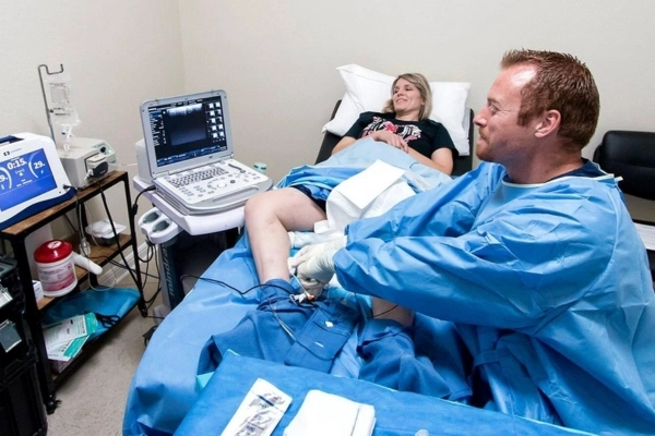
Your Texas Vein Care Team
Meet the physicians who evaluate venous reflux and recommend care after reviewing symptoms, examination findings, and imaging when appropriate.
Our Locations
Find a Texas Vein Experts clinic near you in Dallas–Fort Worth, Houston or San Antonio. Open a region to see every location.

Dallas–Fort Worth
9 clinics
Houston
6 clinics
San Antonio
4 clinicsCommon Questions
Clear answers to common patient questions about possible causes, when to get checked, vein evaluation, and treatment options.
Varicose veins are enlarged superficial veins that may become raised, twisted, or rope-like. They can develop when vein walls stretch or one-way valves no longer keep blood moving efficiently upward, allowing pressure to build in the vein.
A common mechanism is valve dysfunction and venous reflux, which allow blood to move backward and collect in superficial veins. Family history, age, pregnancy, prolonged standing or sitting, and other factors can increase the likelihood of developing them.
Not always. Some people have visible veins with little or no discomfort, while others have aching, heaviness, swelling, itching, tenderness, or skin changes. Evaluation can help separate an appearance-only concern from symptomatic venous disease.
Varicose veins are generally larger and may be raised or bulging. Spider veins are much smaller, flatter, and web-like. Reticular veins fall between them and often appear as flat blue or green feeder veins beneath the skin.
They can occur with aching, heaviness, throbbing, cramping, itching, tenderness, or swelling. These symptoms also have non-venous causes, so the appearance of a varicose vein alone does not identify the source of every leg symptom.
An established enlarged varicose vein usually does not simply return to a normal vein on its own. Symptoms can fluctuate, and pregnancy-related veins may change after delivery, but persistent veins should be evaluated if they are causing concern.
Movement, calf-muscle activity, elevation, and weight management may help some people manage venous symptoms. These measures do not physically eliminate an established varicose vein.
Compression can be useful for symptom management in selected patients, but it is not the right long-term strategy for everyone and should not be presented as a universal prerequisite. Recommendations depend on the clinical situation, treatment plan, and insurance requirements.
Duplex ultrasound can show blood-flow direction, valve function, reflux, vein anatomy, and other findings that help determine whether visible varicose veins connect to an underlying refluxing source vein.
The choice depends on which vein is abnormal, where reflux begins, the size and shape of the target vein, symptoms, medical history, and ultrasound findings. One patient may need more than one technique or a staged plan.
The four treatment pathways represented in this wireframe are radiofrequency ablation, VenaSeal, Varithena, and compounded sclerotherapy. Each serves a different role depending on vein anatomy and the treatment objective.
Yes. Treatment addresses selected veins that are abnormal at the time of care, but it cannot guarantee that other veins will never become varicose later. Follow-up can address new or residual concerns if they develop.
Pregnancy can increase venous pressure and make varicose veins more likely to appear. Management and the timing of procedures are individualized, so pregnancy-related concerns should be discussed with the appropriate clinician rather than treated as a routine procedural indication.
Coverage depends on the insurance plan and whether symptoms, clinical findings, ultrasound results, and other medical-necessity requirements are met. Treatment performed only for appearance may be considered cosmetic.
 English English
 Spanish